Pleural Mesothelioma Ct - Malignant Pleural Mesothelioma Presenting With A Giant Posterior Mediastinal Mass Consultant360 / Malignant pleural mesothelioma (mpm) is a highly aggressive malignant tumor that arises from mesothelial cells of pleural cavity.
Pleural Mesothelioma Ct - Malignant Pleural Mesothelioma Presenting With A Giant Posterior Mediastinal Mass Consultant360 / Malignant pleural mesothelioma (mpm) is a highly aggressive malignant tumor that arises from mesothelial cells of pleural cavity.. Although the chest film findings of pleural mesothelioma are well described, there are few descriptions of the findings of computed tomography (ct). One study showed pleural thickening was evident in 94% of pleural mesothelioma patients who underwent a ct scan. These fibers get lodged into the protective lining of the lungs (the pleura), causing genetic mutations in the surrounding cells. This lets the doctor compare areas of higher radioactivity on the pet. Doctors also use mesothelioma blood tests to measure treatment response.
Although the chest film findings of pleural mesothelioma are well described, there are few descriptions of the findings of computed tomography (ct). The final stages of pleural mesothelioma involve chest pain, shortness of breath. Clinically active cancer other than mesothelioma within 5 years prior to start of study treatment; Tumors are still in the linings of the lung (pleura) but haven't yet spread to the lungs. The main risk factor for mpm is asbestos exposure with most cases discovered in elderly males after a long latency period.
Multimodality Imaging For Characterization Classification And Staging Of Malignant Pleural Mesothelioma Radiographics from pubs.rsna.org European journal of nuclear medicine and molecular imaging, 2014. Malignant pleural mesothelioma (mpm) is a highly aggressive malignant tumor that arises from mesothelial cells of pleural cavity. 37 full pdfs related to this paper. ct scanning not reliable for correct diagnosis of pleural mesothelioma, says new study. (9 days ago) increasingly, fdg pet/ct is used for diagnosis and management of malignant pleural mesothelioma. Their tests on 315 patients have revealed that ct. mesothelioma is a malignant neoplasm originating from pleural or peritoneal surfaces; A ct (or cat) scan or an mri is usually performed.
Although it is less effective at detecting peritoneal (abdominal) mesothelioma, ct scans are still the most useful imaging study for diagnosing peritoneal mesothelioma.
ct scanning not reliable for correct diagnosis of pleural mesothelioma, says new study. A short summary of this paper. This lets the doctor compare areas of higher radioactivity on the pet. pleural effusion occurred in 74% of those patients. pleural mesothelioma affects up to 40,000 people worldwide, 1 yet with around 2000 annually in the united states, it is considered a rare cancer. One study showed pleural thickening was evident in 94% of pleural mesothelioma patients who underwent a ct scan. Although the chest film findings of pleural mesothelioma are well described, there are few descriptions of the findings of computed tomography (ct). More common than mesothelioma occurring in the abdomen. A biopsy is the only definitive way to confirm a mesothelioma diagnosis. Some machines can do both a pet and ct scan at the same time. Comparison of ct and mr imaging. Although it is less effective at detecting peritoneal (abdominal) mesothelioma, ct scans are still the most useful imaging study for diagnosing peritoneal mesothelioma. 37 full pdfs related to this paper.
Four hundred and nine patients with malignant pleural mesothelioma, from 76 centres in the uk and 2 in australia, were randomly assigned to asc alone (n=136); If a large amount of fluid is present, abnormal cells may be detected by cytopathology if this fluid is aspirated with a syringe. Clinically active cancer other than mesothelioma within 5 years prior to start of study treatment; Staging of malignant pleural mesothelioma: Although, ct has intrinsic limitations due to low soft tissue contrast and the current staging system as well as criteria for evaluating response, it does not consider the complex growth pattern of this tumor.
References In Morphologic And Functional Imaging Of Malignant Pleural Mesothelioma European Journal Of Radiology from els-jbs-prod-cdn.jbs.elsevierhealth.com 2 although almost always associated with asbestos exposure, a careful occupational, leisure, travel, and residential history may be needed to uncover exposure sources to access legal compensation. Comparison of ct and mr imaging. 37 full pdfs related to this paper. One study showed pleural thickening was evident in 94% of pleural mesothelioma patients who underwent a ct scan. The investigators anticipate that the intrapleural of the vaccine strain measles virus will enable the virus to specifically infect and kill cancer cells and. Diagnosis of malignant pleural mesothelioma is difficult because its early symptoms are commonly persistent cough, trouble catching your. 2 department of radiology, dongguk university ilsan. It is the least advanced stage.
ct is the first imaging technique used for diagnosis, staging, and assessment of therapy response.
pleural mesothelioma can cause fluid to build up around the lungs in the chest (called a pleural effusion). The 2016 mesothelioma audit data reported that in the uk in 2014 pleural mesothelioma accounted for 2179 cases (97%), with 70 peritoneal cases (approximately 3%).1 in 2007, the british thoracic society (bts) statement on mesothelioma was published in response to a request from the About 75 percent of all mesothelioma cases diagnosed each year are pleural mesothelioma. It is the least advanced stage. pleural mesothelioma commonly presents with dyspnea. ct is particularly valuable in assessing the extent of malignant mesothelioma as well as establishing the presence of pleural effusion or parenchymal disease obscured by a pleural effusion. Doctors also use mesothelioma blood tests to measure treatment response. Although, ct has intrinsic limitations due to low soft tissue contrast and the current staging system as well as criteria for evaluating response, it does not consider the complex growth pattern of this tumor. Pleura) and chest wall (the "parietal" Unlike other malignant mesotheliomas that occur in the abdomen, heart, or testicles, this form of mesothelioma is a cancer that occurs in the pleura, or the tissue lining of the chest cavity that encloses the lungs. Computed tomography features in malignant pleural mesothelioma and other commonly seen pleural diseases. Staging of malignant pleural mesothelioma: The investigators anticipate that the intrapleural of the vaccine strain measles virus will enable the virus to specifically infect and kill cancer cells and.
Stage 1 mesothelioma is the first of four malignant pleural mesothelioma stages. If a large amount of fluid is present, abnormal cells may be detected by cytopathology if this fluid is aspirated with a syringe. Computerized tomography (ct scan) of the chest or abdominal region, along with magnetic resonance imaging or mri, and tissue biopsy of the tissue that makes up the lining of the lungs are some of the tests that a doctor usually runs to check for mesothelioma. Their tests on 315 patients have revealed that ct. Ipsilateral pleural effusion/ hydropneumothorax • ct chest:
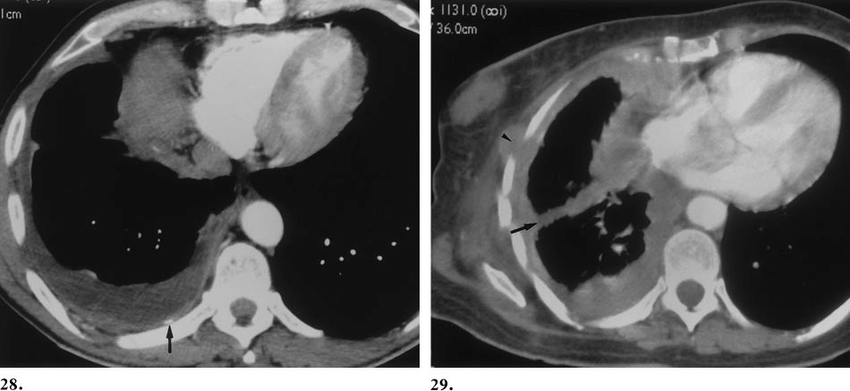
29 28 Axial Ct Scan Of A Patient With A Right Sided Mesothelioma Download Scientific Diagram from www.researchgate.net Mirvis s, dutcher jp, haney pj, et al. European journal of nuclear medicine and molecular imaging, 2014. Asbestosis is often detected through ct scans. ct of malignant pleural mesothelioma. pleural mesothelioma can cause fluid to build up around the lungs in the chest (called a pleural effusion). This retrospective study aimed to investigate the prognostic value of the suvmax in patients with mpm. Wang zj, reddy gp, gotway mb, et al. The tumor originates from cells of the visceral or parietal pleural and is linked to asbestos exposure with a median latency of 44.6 years .due to the latency between exposure and onset of mesothelioma and the ongoing use of asbestos in parts of the world, the incidence is expected to rise.
This phase i clinical trial investigates the side effects and the best dose of local (intrapleural measles virus therapy in treating patients with malignant pleural mesothelioma (mpm).
European journal of nuclear medicine and molecular imaging, 2014. A ct scan is a radiographic technique. pleural mesothelioma is a type of cancer caused by asbestos fibers becoming embedded in the lining of the lungs. This report describes the ct findings in five cases of pleural mesothelioma. @article{osti_5950539, title = {ct findings in malignant pleural mesothelioma related to nonoccupational exposure to asbestos and fibrous zeolite (erionite)}, author = {erzen, c and eryilmaz, m and kalyoncu, f and bilir, n and sahin, a and baris, y i}, abstractnote = {endemic malignant pleural mesothelioma (mpm) in turkey is related to two mineral fibers, tremolite asbestos and fibrous zeolite. pleural mesothelioma commonly presents with dyspnea. Heelan rt, rusch vw, begg cb, et al. The main risk factor for mpm is asbestos exposure with most cases discovered in elderly males after a long latency period. About 75 percent of all mesothelioma cases diagnosed each year are pleural mesothelioma. Malignant pleural mesothelioma (mpm) is an aggressive thoracic malignancy with a dismal prognosis. J comput assist tomogr 1983; The only known cause of pleural mesothelioma is genetic damage caused by exposure to asbestos fibers. Centrally located tumors with radiographic evidence (ct or mri) of local invasion of major blood vessels;


0 Comments